How the amyloid beta cascade may lead to neurological damage
In people with AD, the presence of amyloid beta is believed to trigger a pathological cascade that may promote the abnormal phosphorylation of tau protein, which leads to the formation of neurofibrillary tangles within neurons.1,2
Together, abnormal accumulation of amyloid beta plaques and neurofibrillary tangles can impair neuronal function and may cause neurodegeneration (atrophy).1,3
These pathological changes of Alzheimer’s disease are thought to begin decades before any clinical symptoms appear.²
Pathological Changes Across Alzheimer's Disease Stages4
To learn more about a specific disease stage, use the tappableclickable symbols.
A (Amyloid Beta Accumulation)
It is believed that amyloid beta accumulation starts decades before the onset of clinical symptoms, setting off a cascade that leads to the formation of neurofibrillary tangles and the initiation of neurodegeneration in Alzheimer’s disease.4
Synaptic Dysfunction
When amyloid beta is no longer cleared from the brain properly, it accumulates and is widely believed to lead to synaptic dysfunction and neurodegeneration long before the first symptoms of Alzheimer’s disease start to become visible.4
Tau-mediated Neuronal Injury
The accumulation of amyloid beta is believed to promote the abnormal phosphorylation of tau protein, which leads to the formation of neurofibrillary tangles within neurons.4
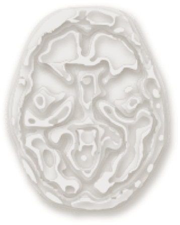
Brain Structure
Together, abnormal accumulation of amyloid beta plaques and neurofibrillary tangles may cause neurodegeneration through neuronal and synaptic loss (atrophy).4
Cognition
The MCI stage of AD may show evidence of disease pathology along with impairment in 1 or more cognitive domains that do not interfere with daily functioning.7
Clinical Function
Initially, amyloid beta plaques accumulate in the centers of higher mental function and those related to learning and memory. Ultimately, plaques compromise areas that regulate attention, emotion, and various other activities, severely impacting daily life.8,9
*Mild cognitive impairment.
Adapted from Jack CR Jr, Knopman DS, Jagust WJ, et al. Hypothetical model of dynamic biomarkers of the Alzheimer’s pathological cascade. Lancet Neurol. 2010;9(1):119-128, with permission from Elsevier. https://www.sciencedirect.com/journal/the-lancet-neurology.
An opportunity to diagnose
According to the cascade hypothesis, amyloid beta accumulation is the earliest marker of Alzheimer's disease pathology, occurring upstream of synaptic dysfunction and tau-mediated neuronal injury.5
This makes biomarker confirmation via amyloid positron emission tomography (PET) scan or cerebrospinal fluid (CSF) test all the more important. The diagnostic value of AD as the cause of MCI provides the clinician an opportunity to take action before greater neuronal damage occurs and more cognition and function are lost.6