1991
NYU
During the MCI stage, clinicians may be able to detect very early features of AD that are distinct from other causes of memory loss or other forms of cognitive impairment.2,3
These features can be detected using validated tools such as Mini-Cog, General Practitioner Assessment of Cognition (GPCOG), Memory Impairment Screen (MIS), and Montreal Cognitive Assessment (MoCA).3,4
The AD continuum has evolved over time to include not only dementia, but also MCI and preclinical Alzheimer’s disease. In fact, the concept of MCI as a stage of AD has been accepted for three decades. Today, multiple sets of criteria refer to the MCI stage of AD based on the same defining elements. And in 2018, the FDA issued guidance on the development of drugs focused on early AD treatment.
Tap the ![]() to learn about important events related to the MCI stage of AD.
to learn about important events related to the MCI stage of AD.
Click the ![]() to learn about important events related to the MCI stage of AD.
to learn about important events related to the MCI stage of AD.
NYU
Mayo
Clinic
Key
Symposium
NIA-AA
DSM-5 &
IWG
NIA-AA
& FDA
1991
1999
2003
2011
2013
2018
NYU=New York University; NIA-AA=National Institute on Aging–Alzheimer's Association; DSM-5=Diagnostic and Statistical Manual of Mental Disorders, Fifth Edition; IWG=International Working Group; FDA=Food and Drug Administration.
1991
NYU
Researchers at NYU include MCI among the six disease stages of the global deterioration scale defining cognitive impairment and dementia.5
1999
Mayo Clinic
The Mayo Clinic creates diagnostic criteria to describe patients with early cognitive dysfunction, focused on memory disturbance that did not meet the definition of dementia.3,5
2003
Key Symposium
Key Symposium of opinion leaders held in Stockholm, Sweden broadened the classification and recognized the possibility of other etiologies for MCI.3
2011
NIA-AA
The National Institute on Aging and the Alzheimer’s Association (NIA-AA) develop criteria for the AD continuum, adopting the Key Symposium definition while adding rules for the use of AD biomarkers.3
2013
DSM-5 & IWG
2018
NIA-AA & FDA
NYU=New York University; NIA-AA=National Institute on Aging–Alzheimer's Association; DSM-5=Diagnostic and Statistical Manual of Mental Disorders, Fifth Edition; IWG=International Working Group; FDA=Food and Drug Administration.
In one study of patients with amyloid beta–positive (Aβ+) MCI, the median time to progression was estimated to be 2 years:
PET imaging and CSF tests can determine whether amyloid beta (Aβ) and/or tau pathology is present in the brain. These biomarkers are currently considered valid proxies for neuropathologic changes of AD and are included in the ATN classification system. Recent recommendations by the International Working Group (IWG) require both clinical and biological diagnosis of AD. Although Aβ and tau biomarkers are not sufficient to predict progression or identify a stage of AD on their own, they are necessary to make an AD diagnosis.
ATN=amyloid beta, tau, neurodegeneration.
The IDEAS Study recently showed how AD biomarkers changed the management of patients with MCI or dementia of uncertain etiology. In this longitudinal study published by the Journal of the American Medical Association, the primary endpoint was change in patient management, including AD therapy, other drug therapy, or safety counseling/future planning (target: 30% composite change in each group) after amyloid PET scanning.10
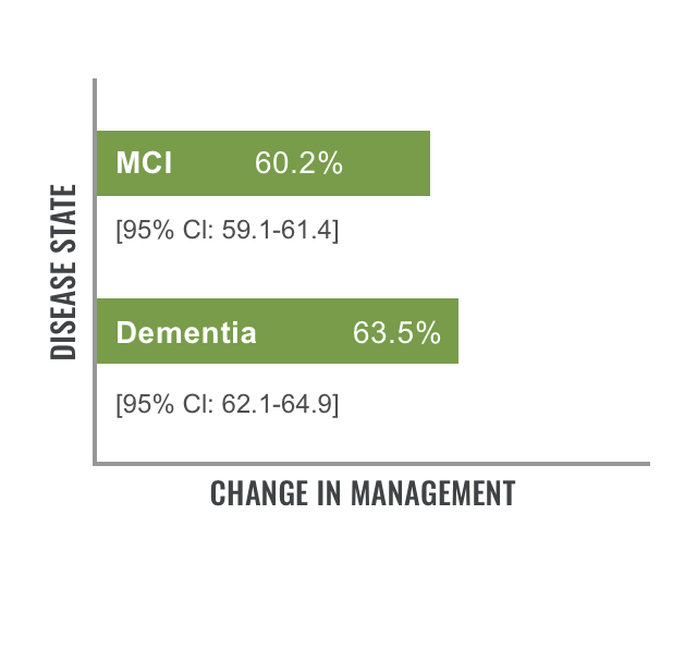
N=11,409; P<0.001.
Early and accurate diagnosis of Alzheimer's disease is important for patient care. Identifying biomarkers may serve as the mechanism to improve early and accurate diagnosis.11,12
 Journal Feature
Learn more about how amyloid PET influenced changes in the management of MCI or dementia.
Read: Rabinovici GD, Gatsonis C, Apgar C, et al. Association of amyloid positron emission tomography with
subsequent change in clinical management among Medicare beneficiaries with mild cognitive impairment or
dementia. 𝘑𝘈𝘔𝘈. 2019;321(13):1286-1294.
Read: Rabinovici GD, Gatsonis C, Apgar C, et al. Association of amyloid positron emission tomography with subsequent change in clinical management among Medicare beneficiaries with mild cognitive impairment or dementia. JAMA. 2019;321(13):1286-1294.